Medical data visualization
Enhance your medical solution, device, or product with advanced medical data visualization.
We understand the critical role that data visualization plays in medicine. Visualization is crucial in particular, for clinical diagnosis, treatment planning, and image analysis. With our expertise, we help you explore medical imaging data in new ways, uncover hidden insights, streamline diagnostics, and also improve treatment planning.
From 3D imaging to bespoke visual solutions, we turn medical data into a powerful tool for better healthcare outcomes.
Leverage the benefits of medical data visualization custom solution
New imaging biomarkers
The interpretation of images can be challenging because they contain more information than you may think. Visualizing patient data can make the process simpler and uncover new biomarkers, providing both morphological and functional insights. In particular, these biomarkers can help detect diseases at earlier stages. What’s more, sometimes even before clinical symptoms appear.
Clinical trials and research
Visual representation of clinical data is an effective tool for investigation and also for making well-informed decisions in clinical trials. What’s more data visualization methods based on AI-enabled medical image segmentation and processing improve the quality of data (higher objectivity, lower inter-observer variability and decrease of required sample size), resulting in cost reduction.

Medical education
Advanced data visualization plays an important role in medical education, particularly in the areas of anatomy, radiology, and surgery. It is widely used in surgical training, including the use of simulators with both physical and virtual 3D medical models. Visual and interactive data presentations can be examined, analyzed, and also transformed into useful knowledge or models.

Easier and faster diagnosis
3D/4D models provide a better understanding of anatomical structures. AI technologies can identify and separate entire objects such as organs and tumors, and their parts (e.g. the aorta, coronary arteries, plaques, and also valves). Surely, this technique enables more accurate examination, reduces the time needed, and enhances consistency among different observers.

Treatment planning
Some new biomarkers become key parameters and help choose the best treatment (e.g. pharmacotherapy, minimally-invasive intervention, surgical treatment) and optimal intervention timing. They play a vital role in treatment planning including radiation or invasive intervention (e.g. implant size, 3D printed personalized implant), enabling better risk assessment.

Treatment evaluation
Visualization methods improve the understanding of data and enhance the assessment of treatment effectiveness. Moreover, they allow for an accurate evaluation of potential complications and early detection of relapse by comparing follow-up studies. These techniques provide a view of changes over time, helping make decisions and improving patient outcomes.
Medical data visualization: selected works

Cinematic volume rendering
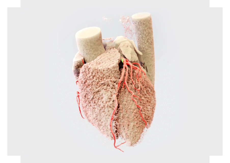
Cinematic volume rendering is a popular data visualization technique, thus often used to present three-dimensional images (3D) of computed tomography.
An example shows how semantic segmentation helps to analyze images (3D) of computed tomography images.
In order that masking can be used to remove the heart, aorta and also coronary arteries.

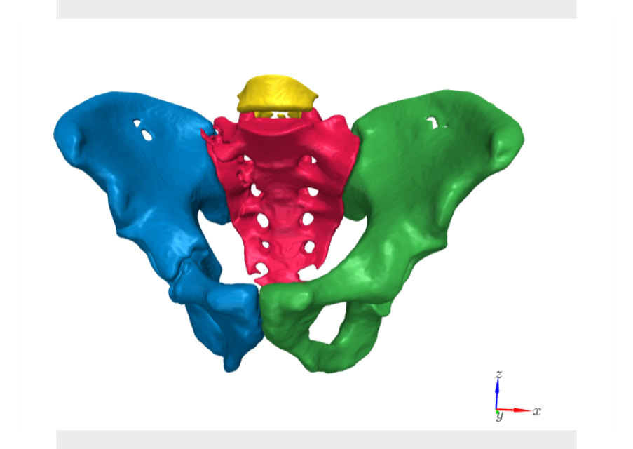
Visualization of dividing the pelvic area and spine into areas
Visualization of the effect of dividing the pelvic area and spine into 4 areas (sacrum, left ilium, right ilium, and also lumbar spine).
Imaging takes into account different types of fractures (fracture, fragmentation and also displacement).

Unveiling the hidden insights
Healthcare data visualization is one of the key stages of data analysis. It enables faster interpretation and a deeper understanding of the hidden patterns. However, raw data often lacks the clarity needed for informed decision-making. This is where the power of artificial intelligence comes into play. By transforming complex data sets into visually compelling representations, we can, consequently, uncover hidden patterns and insights.
Our team of experts specializes in harnessing the potential of medical image visualization. Besides, we develop bespoke solutions that meet the specific needs of our clients. Surely, with us, you can create dedicated solutions that:
- Identify anomalies: help detect anomalies and potential health risks that might be overlooked in traditional analysis
- Provide support in diagnosis: your solution, in essence, will aid clinicians in making diagnoses
- Optimize treatment planning: enable personalized treatment plans based on visual data
- Clarify complex structures: bespoke solutions created by us will provide as a result an understanding of complex details of anatomy
Our medical imaging solutions can streamline your workflows and improve efficiency and accuracy through automated data analysis. Additionally, the transformative power of medical visualization lies in its ability to reveal the hidden intricacies of the human body.
In summary, by visualizing data you can gain deeper insights into anatomical structures.
3D visualization
This techniques have revolutionized surgical planning. Surgeons can now create detailed 3D models of a patient’s anatomy. Moreover, it allows them to simulate procedures before entering the operating room, potentially reducing surgical risks.
